Prostate Cancer Care

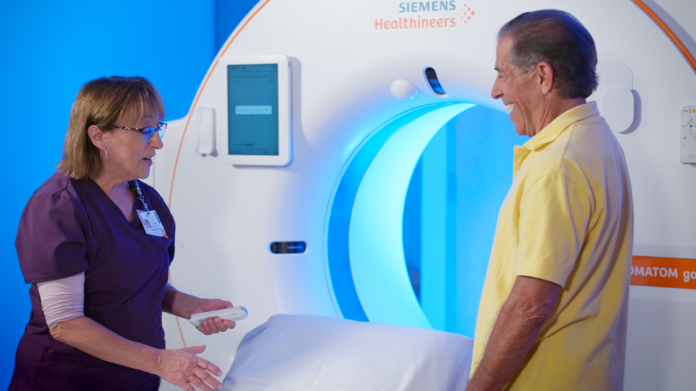
The Center for Cancer Care at Griffin Hospital specializes in diagnosing and treating prostate cancer with state-of-the-art biopsy techniques and sophisticated high-level treatments including Prostate Radiosurgery. We offer advanced treatment for all stages of prostate cancer in a tranquil, healing environment.
In keeping with Griffin’s commitment to providing exceptional, person-centered care, we have highly trained Urologic Surgeons, Medical Oncologists, and Radiation Oncologists who work as a team to provide a multi-disciplinary approach for all newly diagnosed patients.
Contact us at 203-732-1280 for Exceptional, Patient-Centered Prostate Cancer Care
DIAGNOSIS
Prostate screening can find cancer before it causes symptoms. The two most common screening methods for prostate cancer are the prostate-specific antigen (PSA) test and a digital rectal exam.
PSA Test
PSA is a protein made by the prostate. It is made by healthy and cancerous prostate cells. The PSA test measures the level of PSA in the blood. If the PSA level is slightly elevated, but there are no other reasons to suspect prostate cancer, it may only require ongoing monitoring. If the PSA level is too high, has risen significantly, or if a prostate lump is found during a digital rectal exam, other tests may be scheduled, such as a prostate biopsy.
Digital Rectal Exam
The prostate sits near the rectum. This allows clinicians to conduct a digital rectal exam to feel the prostate through the wall of the rectum. During this test, the clinician inserts a gloved finger into the rectum and uses gentle pressure to feel for abnormalities in the prostate.
Prostate Biopsy
A prostate biopsy is the removal of a tissue from the prostate gland, which are then sent to a laboratory for testing. A prostate biopsy is conducted if part of the prostate looks suspicious or after abnormal results from a PSA test or digital rectal exam. A prostate biopsy is the definitive way to find out if there are cancer cells in the prostate.
UroNav® Prostate Fusion Biopsy
At the Center for Cancer Care at Griffin Health, patients can have a prostate biopsy using the UroNav Prostate Fusion Biopsy. This advanced imaging system combines pre-biopsy images from magnetic resonance imaging (MRI) with ultrasound-guided biopsy images in real time to create a detailed, three-dimensional view of the prostate. An improvement to a standard biopsy, this imaging system is a breakthrough improvement to a standard prostate biopsy as it can:
- Accurately pinpoint suspicious areas
- Reduce the number of tissue samples required during biops
- Reduce the risk of infection, bleeding and pain
- Shorten recovery after biopsy

Prostate Specific Membrane Antigen (PSMA) PET/CT Imaging
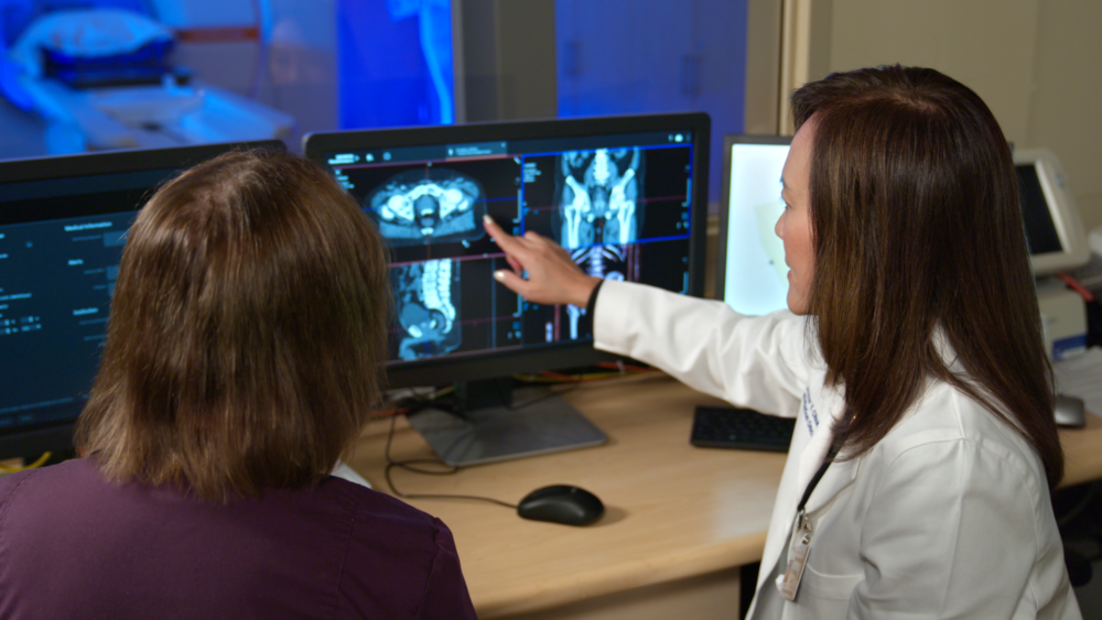
In addition to state-of-the-art imaging for more precise biopsies, Griffin is offering a revolutionary new imaging technique to find prostate cancer outside of the prostate with greater accuracy and far earlier than older imaging methods. The Prostate Specific Membrane Antigen (PSMA) PET/CT imaging technology can identify cancer both in and outside the prostate gland and especially benefits men whose cancer has recurred and are at risk for it spreading to other parts of the body, even after previous treatments.
The (PSMA) PET/CT method incorporates a radioactive tracer as a molecular imaging marker for prostate cancer. The tracer is injected an hour before imaging and binds to PSMA, a protein on the surface of prostate cancer cells. Cancerous cells are identified as bright spots on the PET scan, and their location is revealed on a CT scan conducted at the same time. This helps develop targeted treatment plans, which may include radiation, chemotherapy, hormone medications, or surgery, to address our patient’s individual needs.
TREATMENT
At the Center for Cancer Care at Griffin Hospital, we offer the most advanced technologies available in medicine today, including Intensity Modulated Radiation Therapy (IMRT), Image Guided Radiation Therapy (IGRT) and Stereotactic Body Radiation Therapy (SBRT), together with the medical expertise of Yale Therapeutic Radiology, one of the largest and most well respected groups of radiation oncologists in the region.
Intensity Modulated Radiation Therapy (IMRT) with Image-Guided Radiation therapy (IGRT)
When External Beam Radiation Therapy (RT) is recommended to treat localized prostate cancer, the combination of IMRT with IGRT is the most advanced and patient-centered treatment option available. This highly precise technology intricately maps the prostate to safely allow for more effective doses of radiation to be accurately delivered to the cancer while minimizing radiation to normal tissues including the rectum, bladder or bowel.
How Intensity Modulated Radiation Therapy (IMRT) Works
IMRT technology provides optimally precise radiation therapy to the prostate while effectively limiting any adverse effects to surrounding normal structures such as the rectum, bladder or bowel. To achieve this high level of safety, IMRT varies the intensity of radiation beams, allowing stronger doses to reach specific areas of the tumor. The varying intensity can be generated by adjusting the opening of the RT beam (collimator) with a fixed gantry position or by changing the beam opening during an arc.
Treatments are delivered over 5-8 weeks either using standard doses of radiation or hypofractionated doses in which the total dose of radiation is divided into large doses and treatments are given once a day or less often.
How Image Guided Radiation Therapy (IGRT) Works
IGRT provides an accurate location of your prostate during treatment. This technology is vitally important to prostate cancer radiation therapy because the prostate gland can vary in position due variations in the size of the bladder and rectum. This technology tracks the location of the prostate and the surrounding organs before each radiation dose is delivered.
Hydrogel
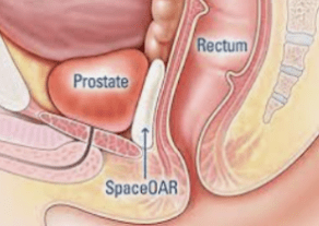 To enhance patient safety, Space OARTM Hydrogel, a soft gel material, can be used to temporarily create a space between the prostate and rectum. This greatly minimizes any potential short-or long-term side effects caused radiation. A urologist can place the gel during an outpatient procedure.
Stereotactic Body Radiation Therapy (SBRT)
Stereotactic Body Radiation Therapy (SBRT) is offered to carefully selected men who are in the early stages of prostate cancer and do not need nodal irradiation. Recommendation for this procedure is based on guidelines from the National Comprehensive Cancer Network (NCCN), American Society for Radiation Oncology (ASTRO) & American Society of Clinical Oncology (ASCO), and American Urological Association (AUA).
How does Stereotactic Body Radiation Therapy (SBRT) Work?
SBRT is an advanced form of radiation therapy in which a full calculated treatment of radiation is administered in 1-5 smaller doses over the course of 5 days. This technology is an improvement over conventional external beam radiation treatments where the total radiation dose this is delivered over several weeks. In order to efficiently administer radiation using stereotactic radiosurgery, Griffin utilizes high-resolution imaging to define exactly where the tumor is in relation to the normal surrounding organs, including lung, bladder, and rectum.
We will need to immobilize your body during this procedure because it allows us to precisely treat the affected area and effectively reproduce the treatment. At Griffin, we build either a custom body cast and use radiopaque markers to establish a system of three-dimensional coordinates for treatment.
We also place tiny, gold seeds that are about the size of a grain of rice called fudicial markers in and/or around a tumor to show exactly where it is in the body.
What to Expect During Your Stereotactic Body Radiation Therapy (SBRT) Treatment
CT Simulation
 After you and your physician have decided that SBRT is best for you, we will need to precisely map your body to ensure the treatment is safe and effective. A specialized type of Computerized Tomography (CT) called a CT Simulator produces 3D images of your internal structure that allow your physician to map out your prostate and surrounding organs such as lymph nodes, rectum, bladder, and bowel. Highly accurate measuring devices are then employed to record the treatment site in relation to external markers.
Treatment Planning
After making a 3D map of your anatomy, your physician, in coordination our highly skilled physics team, will develop a Volumetric Modulated Arc Plan. This plan will specify the doses of radiation needed to treat the tumor.
Daily Treatments
Daily external beam radiation is delivered by our state-of-the-art Elekta Synergy Linear Accelerator. This sophisticated device precisely delivers doses of therapeutic radiation to kill cancer cells. Immediately before each treatment, a Cone-Beam CT or IGRT localization is performed. This allows accurate daily localization of the tumor within millimeters to correct for daily prostate motion. Our experienced Radiation Therapy staff delivers the daily treatments as prescribed by your doctor. A typical treatment will be approximately 15 minutes.
Concerns about Radiation Therapy
Many patients are concerned about the amount of radiation they receive. Radiation treatments for cancer are very safe and effective. The radiation treatments are high energy that travel like a beam of light (without scatter) to destroy cancer cells that have abnormal DNA. This is unlike the radiation used in medical imaging, such as chest X-rays, CT scans, and fluoroscopy dental X-rays, which are low-energy radiation that do not affect cancer cells. However, these types of radiation are not as precise as radiation therapy and can “scatter,” potentially requiring patients to wear shields for their protection.
Follow Up
In conjunction with your doctor, a plan for follow up visits and testing is made after the completion of your treatment. Most patients are seen within three months for a prostate-specific antigen (PSA) blood test. Typically, PSA levels and a digital rectal exams provide the best way to measure the success of the treatments.
Please call 203-732-1280 to Learn More